


The anatomy of the lower extremity venous system is highly variable.Understanding the individual characteristics of the venous system structure plays an important role in evaluating instrumental examination data and selecting the correct treatment approach.
The veins of the lower limbs are divided into superficial veins and deep veins.The superficial venous system of the lower limbs originates from the venous plexus of the toes and forms the venous network of the dorsum of the foot and the cutaneous arch of the dorsum of the foot.The medial and lateral marginal veins arise from it and enter the great and small saphenous veins respectively.The great saphenous vein is the longest vein in the body, containing 5 to 10 pairs of valves, with a normal diameter of 3-5 mm.It originates in the lower third of the leg anterior to the medial epicondyle and ascends into the subcutaneous tissue of the calf and thigh.In the inguinal area, the great saphenous vein empties into the femoral vein.Sometimes the great saphenous vein in the thigh and calf can be represented by two or even three trunks.The lesser saphenous vein starts along the side of the leg in the lower third of the leg.In 25% of cases, it drains into the popliteal vein in the popliteal fossa region.In other cases, the small saphenous vein can ascend above the popliteal fossa and drain into the femoral, great saphenous, or deep thigh veins.
The deep dorsal vein of the foot originates from the dorsal plantar vein and flows into the dorsal venous arch, from where blood flows into the anterior tibial vein.At the level of the upper third of the leg, the anterior and posterior tibial veins merge to form the popliteal vein, located laterally and slightly posterior to the artery of the same name.In the popliteal region, both the small saphenous vein and the knee vein drain into the popliteal vein.The deep thigh veins usually drain into the femoral vein 6-8 cm below the inguinal fold.Above the inguinal ligament, this vessel receives the epigastric vein (the deep vein around the ilium) and enters the external iliac vein, which joins the internal iliac vein at the sacroiliac joint.The paired common iliac veins begin after the union of the external and internal iliac veins.The left and right common iliac veins join to form the inferior vena cava.It is a large container without a valve, 19-20 cm long and 0.2-0.4 cm in diameter.The inferior vena cava has mural and visceral branches, and blood flows from the lower limbs, lower trunk, abdominal organs, and small pelvis.
Perforating (communicating) veins connect deep veins to superficial veins.Most of them have valves located on the fascia through which blood flows from superficial to deep veins.There are direct perforating veins and indirect perforating veins.The direct type directly connects the deep and superficial vein networks, while the indirect type connects indirectly, that is, first flows into the muscular veins and then into the deep veins.
The vast majority of perforating veins arise from tributaries rather than from the main trunk of the great saphenous vein.Perforating veins on the medial surface of the lower third of the leg are incompetent in 90% of patients.In the lower leg, insufficiency of the Cockett perforating vein connecting the posterior branch of the great saphenous vein (Leonardo's vein) to the deep vein is most commonly observed.There are usually 2-4 most permanent perforating veins (Dodd, Gunter) in the middle and lower third of the thigh, which directly connect the great saphenous vein trunk and the femoral vein.With varicose transformation of the small saphenous veins, communicating venous insufficiency is most often observed in the mid-leg, lower third, and lateral malleolus regions.
clinical course of disease
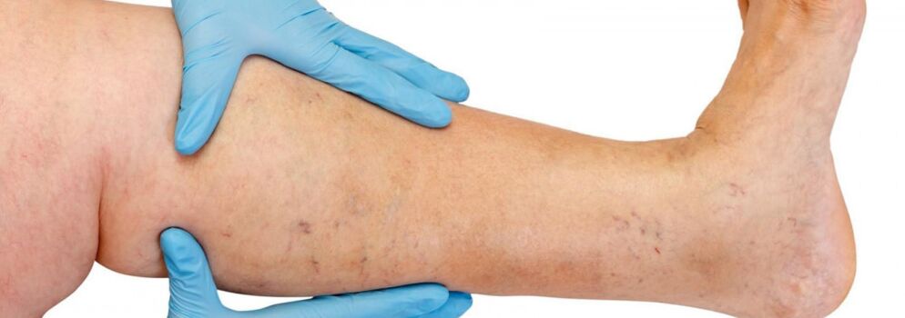
Varicose veins mostly occur in the great saphenous vein system, less commonly in the small saphenous vein system, starting from the tributaries of the venous trunks of the legs.The natural course of the disease in the early stages is quite favorable; for the first 10 years or more, patients may not be bothered by anything other than cosmetic defects.Later, if left untreated, a feeling of heaviness, fatigue and swelling in the legs will begin after physical activity (long walks, standing) or in the afternoon, especially during hot seasons.Most patients complain of leg pain, but detailed questioning reveals that this is the feeling of fullness, heaviness, and fullness in the legs.Even short rest and an elevated limb position can reduce the severity of the sensation.These symptoms are characteristic of venous insufficiency at this stage of the disease.If we are talking about pain, we need to rule out other causes (insufficient arterial blood supply to the lower limbs, acute venous thrombosis, joint pain, etc.).Subsequent disease progression, in addition to an increase in the number and size of dilated veins, leads to the development of nutritional disorders, often due to an increase in incompetent perforating veins and the development of deep venous valvular insufficiency.
If the perforating veins are insufficient, dystrophic disorders are limited to any surface of the leg (lateral, medial, posterior).Nutritional disorders initially manifest as localized hyperpigmentation of the skin, followed by thickening (induration) of subcutaneous adipose tissue until cellulite is formed.The process culminates in the formation of ulcerative necrotic defects that can be 10 cm or larger in diameter and penetrate deep into the fascia.The typical site of venous trophic ulcers is the medial malleolus area, but calf ulcers can be variable and multiple in location.During the dystrophic stage, severe itching and burning sensations occur in the affected area; some patients develop microbial eczema.Pain in the area of the ulcer may not show up, although in some cases the pain can be severe.During this stage of the disease, the legs continue to feel heavy and swollen.
Diagnosis of varicose veins
Diagnosing varicose veins in the preclinical stage is particularly difficult because such patients may not have varicose veins in their legs.
In such patients, the diagnosis of varicose veins in the legs is erroneously rejected despite the presence of symptoms of varicose veins, ultrasound data indicating that the patient has a relative with this disease (genetic predisposition), and initial pathological changes in the venous system.
All of this can lead to missing the deadline for optimal treatment initiation, irreversible changes in the formation of vein walls, and the development of very serious and dangerous complications of varicose veins.Only by identifying the disease at an early preclinical stage is it possible to prevent pathological changes in the venous system of the legs with a minimal therapeutic effect on varicose veins.
Various diagnostic errors can be avoided and the correct diagnosis made only by a thorough examination of the patient by an experienced specialist, by correctly interpreting all the patient's complaints, by analyzing the medical history in detail and by using the most modern equipment (instrumental diagnostic methods) to obtain the maximum possible information about the state of the leg's venous system.
A duplex scan is sometimes performed to determine the exact location of the perforating vein, identifying venous venous return with a color code.If the valve is not fully functional, their valve will stop closing completely during a Valsava maneuver or compression test.Valvular insufficiency results in the development of venous reflux, with high reflux through the incompetent saphenofemoral junction and low reflux through incompetent perforating veins in the leg.Using this method, the reverse flow of blood through the prolapsed leaflets of a dysfunctional valve can be recorded.This is why diagnosis is multi-stage or multi-layered.Under normal circumstances, the diagnosis is made after ultrasound and examination by a phlebologist.But in particularly difficult cases, inspections must be carried out in stages.
- First, a thorough examination and interview by a vein surgeon;
- If necessary, the patient will be sent for other instrumental investigation methods (double vascular scan, venous scintigraphy, lymphoscintigraphy);
- Patients with concomitant diseases (osteochondrosis, varicose eczema, lymphovenous insufficiency) can seek consultation or other research methods from leading expert consultants in these diseases;
- All patients requiring surgery first receive a consultation with the operating surgeon and, if necessary, an anesthetist.
treat
Conservative treatment is mainly suitable for patients who have contraindications for surgical treatment: due to general conditions, slight dilation of the veins only causes cosmetic inconvenience, or who refuse surgical treatment.The goal of conservative treatment is to prevent further progression of the disease.In these cases, the patient should be advised to wrap the affected area with an elastic bandage or wear elastic stockings, regularly place the legs in a horizontal position and perform special exercises for the feet and calves (flexion and extension of the ankle and knee joints) to activate the muscular venous pump.Elastic compression accelerates and enhances the blood flow in the deep veins of the thigh, reduces the blood volume of the great saphenous vein, prevents the formation of edema, improves microcirculation, and helps normalize tissue metabolic processes.Bandaging should be started before getting up in the morning.Apply gentle tension from your toes to your thighs and forcefully grip your heels and ankle joints.Each subsequent round of bandages should overlap the previous round by half.It is recommended to use certified medical knitwear and individually select the degree of compression (1 to 4).Patients should wear comfortable, hard-soled, low-heeled shoes and avoid prolonged standing, strenuous labor, and working in hot and humid areas.If, due to the nature of work activities, the patient must sit for long periods of time, special supports of the required height should be placed under the feet, placing the legs in an elevated position.It is recommended to walk a little or stand on tiptoes 10-15 times every 1-1.5 hours.The resulting contraction of the calf muscles improves blood circulation and increases venous outflow.When sleeping, your legs need to be elevated.
Patients are advised to limit their water and salt intake, normalize their weight, and regularly take diuretics and drugs that improve venous tone.Depending on the indication, drugs that improve tissue microcirculation are prescribed.For treatment, nonsteroidal anti-inflammatory drugs are recommended.
Physical therapy plays an important role in preventing varicose veins.For uncomplicated forms, water procedures are useful, especially swimming, with foot baths in warm (no higher than 35°) water with a solution of 5-10% common salt.
Compression sclerotherapy
The indications for injection treatment of varicose veins (sclerotherapy) are still debated.This method involves introducing a sclerosing agent into the dilated vein, causing it to further compress, relax, and harden.Modern drugs used for these purposes are very safe, that is, they do not cause necrosis of the skin or subcutaneous tissue when administered extravascularly.Some experts use sclerotherapy for almost all forms of varicose veins, while others reject this approach entirely.The truth is likely somewhere in between, and for young women in the early stages of the disease, treatment with injections makes sense.The only problem is that they must be warned of the possibility of recurrence (higher than surgical intervention), the need to continue wearing a fixed compression bandage for a long time (up to 3-6 weeks), and the potential need for multiple treatments to fully harden the vein.
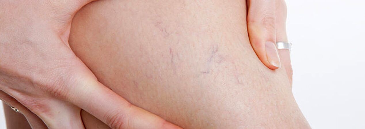
The group of patients with varicose veins should include patients with telangiectasia ("spider veins") and reticular dilatation of the small saphenous veins, as these conditions occur for the same reason.In this case, in addition to sclerotherapy, you canpercutaneous laser coagulation, but only if damage to deep veins and perforating veins is excluded.
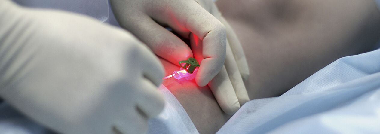
Percutaneous Laser Coagulation (PLC)
This is a method based on the principle of selective photocoagulation (photothermolysis), which is based on the different absorption of laser energy by various substances in the body.A feature of this approach is the contactless nature of the technology.The focusing head concentrates energy into the blood vessels of the skin.Hemoglobin in blood vessels selectively absorbs laser beams of specific wavelengths.Under the action of laser, the endothelium of blood vessels is destroyed, resulting in the adhesion of the blood vessel walls.
The effectiveness of PLK directly depends on the penetration depth of laser radiation: the deeper the vessel, the longer the wavelength should be, so the indications for PLK are rather limited.Microsclerotherapy is most effective for blood vessels larger than 1.0-1.5 mm in diameter.Considering that spider veins in the legs are widely distributed, have many branches, and have variable blood vessel diameters, a combination treatment method is currently actively used: in the first stage, sclerotherapy is performed on veins with a diameter greater than 0.5 mm, and then laser is used to remove the remaining "stars" with smaller diameters.
The procedure is virtually painless and safe (no skin cooling or anesthesia is used) because the device's light falls into the visible part of the spectrum, and the light wavelengths are designed so that water in the tissue does not boil and the patient does not get burned.For patients with high pain sensitivity, it is recommended to apply a local anesthetic ointment beforehand.Redness and swelling should subside within 1-2 days.After treatment, some patients may experience darkening or lightening of the skin in the treated area for approximately two weeks, which then disappears.For people with fair skin, this change is barely noticeable, but for patients with dark skin or tan, the risk of this temporary pigmentation is quite high.
The number of surgeries depends on the complexity of the case - the blood vessels are located at different depths and the lesions may be small or occupy a considerable area of skin surface, but usually no more than four laser treatments (5-10 minutes each) are required.Because the device's light pulses have a unique "square" shape, they are able to achieve maximum results in such a short period of time; this increases their effectiveness compared to other devices and also reduces the likelihood of side effects after surgery.
surgical treatment
Surgery is the only radical treatment for patients with varicose veins in the lower limbs.The goal of surgery is to eliminate the causative mechanism (veno-venous reflux).This is accomplished by resection of the main trunks of the great and small saphenous veins and ligation of the incompetent communicating veins.
Surgical treatment of varicose veins has a history of more than 100 years.Previously, many surgeons still did this, making large incisions along the varicose veins and using general or spinal anesthesia.The scars left after this "mini-phlebectomy" remain a lifelong reminder of the surgery.The first venous surgery (according to Madelon, according to Schade) was so traumatic that its harm outweighed the dangers of varicose veins.
In 1908, the American surgeon Babcock proposed a method of subcutaneous vein retraction using a rigid metal probe with an olive.After modifications, this surgical method of removing varicose veins is still used in many public hospitals.Varicose veins are removed using a separate incision, as recommended by Surgeon Narat.Therefore, the classic phlebectomy is known as the Babcock-Narat method.According to Babcock-Narat, phlebectomy has disadvantages - a large scar is left after the procedure and skin sensitivity is compromised.Reduced work ability for 2-4 weeks makes it difficult for patients to agree to surgical treatment of varicose veins.
Phlebologists have developed a unique technique that can treat varicose veins in just one day.Operation in complex situationsCombination technology.The main main trunk of large varicose veins is removed by inversion peeling with minimal intervention through a small skin incision (2 to 7 mm), leaving virtually no scarring.The use of minimally invasive techniques involves minimal tissue trauma.The result of this procedure is the elimination of varicose veins with excellent cosmetic results.Combined surgical treatment is performed under total intravenous or spinal anesthesia, with a maximum hospital stay of up to 1 day.
Surgical treatment includes:
- Cross resection - across the trunk of the great saphenous vein where it empties into the deep venous system;
- Dissection is the removal of varicose vein fragments.Only the varicose veins are removed, not the entire vein (as in the classic version).
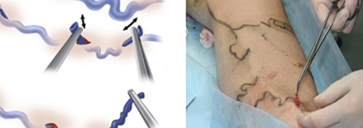
actuallyvenous resectionReplaced the Narat technique to remove varicose tributaries of the main veins.Previously, a skin incision of 1-2 cm to 5-6 cm is made along the course of the varicose vein, through which the vein is separated and excised.The desire to improve the cosmetic results of the intervention and to be able to remove the vein not through a traditional incision but through a small incision (puncture) has forced doctors to develop tools that allow them to accomplish almost the same thing through the smallest skin defect.This is where phlebectomy "hooks" of various sizes and configurations come in, as well as special spatulas.And start using a scalpel with a very narrow blade or a needle with a fairly large diameter to pierce the skin (for example, a needle used to draw venous blood for analysis, with a diameter of 18G).Ideally, the puncture mark with such a needle will be almost invisible after a while.
Some forms of varicose veins are treated on an outpatient basis under local anesthesia.The minimally invasive procedure of venous resection and the low risk of intervention allow the procedure to be performed in day hospitals.After minimal observation in the clinic after surgery, patients can go home on their own.During the postoperative period, maintain an active lifestyle and encourage active walking.The period of temporary incapacity usually does not exceed 7 days, after which work can be resumed.
When is microphlebectomy used?
- When the diameter of the great saphenous vein or small saphenous vein varicose trunk is greater than 10 mm;
- After thrombophlebitis occurs in the main subcutaneous trunk;
- Trunk recanalization after other types of treatment (EVLT, sclerotherapy);
- Resection of very large individual varicose veins.
It can be a stand-alone procedure or part of a combined varicose vein treatment that combines vein laser treatment and sclerotherapy.The strategy of use is determined individually, always taking into account the results of the ultrasound duplex scan of the patient's venous system.Microphlebotomies are used to remove veins in different areas that have been altered for a variety of reasons, including veins on the face.Professor Varady from Frankfurt developed his convenient instrument and formulated the basic assumptions of modern venectomy.Varadi phlebectomy provides excellent cosmetic results without pain and without hospitalization.It's very hard work, almost jewelry work.
after venous surgery
The usual postoperative period after a "classic" phlebectomy is quite painful.Sometimes a large hematoma occurs and swelling occurs.Wound healing depends on the phlebologist's surgical technique; lymphatic leakage sometimes occurs, with long-term visible scarring; often, there is still a loss of sensitivity in the heel area after a major phlebectomy.
In contrast, after venous resection, the wound does not require suturing, as these are merely punctures, without pain, and no damage to the cutaneous nerves has been observed in practice.However, this result of phlebectomy can only be achieved by very experienced phlebologists.